Real-time Functional Imaging and Neurofeedback
Feb. 12-13, 2015
Gainesville, FL
Niels Birbaumer, University of Tuebingen
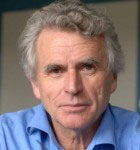 Niels Birbaumer is Professor and Director of the Institute of Medical Psychology and Behavioral Neurobiology, University of Tübingen, Faculty of Medicine as well as the director of the Magnetoencephalography (MEG)-Center. Professor Birbaumer obtained his Ph.D. degree in psychology from the University of Vienna in 1969. His research interests are wide-ranging. Among other things he is dealing with neuronal plasticity and learning, with aspects of epilepsy, of Parkinson's disease and pain disorders. Prof. Birbaumer also conducts research on brain-computer interfaces which should make it possible to exchange information without the use of the limb between the brain and machines. This research is intended as patients with end-stage amyotrophic lateral sclerosis (ALS) allow to communicate in spite of complete paralysis with their environment.
Eberhard Fetz, University of Washington
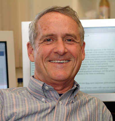 Eberhard Fetz received his B.S. in physics from the Rensselaer Polytechnic Institute in 1961, and his Ph.D. in physics from the Massachusetts Institute of Technology in 1967. He came to the University of Washington for postdoctoral work in neuroscience and has been on the faculty ever since. He is currently Professor in the Department of Physiology & Biophysics, Adjunct Professor in Bioengineering, Affiliate Professor in DXARTS and Head of the Neuroscience Division of the Washington National Primate Research Center. His overall research has concerned the neural control of limb movement in primates. He pioneered the recording of spinal interneurons in behaving monkeys and showed that spinal neurons share many properties of cortical cells, including preparatory activity prior to instructed movements. Most recently, his lab has developed an implantable recurrent brain-computer interface, called Neurochip that can record activity of cortical cells during free behavior and convert this activity in real time to stimulation of cortex, spinal cord or muscles.
Abstract:
Volitional Control of Neural Activity: Past and Future
Eberhard E. Fetz, Ph.D.
Department of Physiology & Biophysics, University of Washington, Seattle, WA, USA
Volitional control of neural activity has been demonstrated by experiments involving operant conditioning with neurofeedback, and by brain-computer interfaces involving neural control of external devices. Operant conditioning has allowed the relationships between multiple neurons to be probed directly in non-human primates, and recent experiments have shown that even synaptically connected neurons could be activated quite independently. Delivery of intracranial reinforcement with an autonomous closed-loop brain-computer interface has enabled operant conditioning of neural and muscular activity during free behavior, and opens the way toward testing volitional control of spatiotemporal firing patterns. In human subjects learning to control cursor movement with electrocorticograpic signals recorded from specific cortical sites, the activity at other sites showed strongly modulated activity that changed with learning. This talk will review these and related studies and speculate on future research directions.
Jeffrey Fitzsimmons, University of Florida
D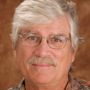 r. Fitzsimmons is a Professor of Radiology at the University of Florida. As a graduate student in neuropsychology at UF he helped design components of the Visual Prosthesis project which mapped the primate visual cortex using cortically stimulated visual phosphenes. Upon graduation (PhD, 1974) he joined the UF Radiology department where he worked with Drs. Scott and Andrew to convert a small bore NMR spectrometer into a prototype MR imaging system. Later, when one of the first 0.15T whole body magnets became available, he began designing radio frequency (RF) coil technology for human brain and spine imaging. For more than ten years he directed the MR Instrumentation core of an NIH funded High Field NMR Research Resource. As available MR field strength increased he led the effort to acquire the first 3T whole body MRI unit for the UF/VA hospitals and developed a series of RF coil arrays for functional brain imaging (fMRI) and other applications. When fMRI was need for NIH funded research with Dr. Lang, he directed the development of one of the first MR compatible fMRI stimulus presentation systems. In the early 1990's (using SBIR funding) he co-founded Applied Resonance Technology (ART) which later merged with MRI Devices to produce a commercial line of RF coil arrays for high field MRI. He also served on the board of MRI Devices and helped negotiate the sale of MRI Devices to IGC in 2004 which was later sold to Philips later the
same year. Dr. Fitzsimmons has been an Angel investor screening over 100 start up companies in the past 10 years and now serves as a Mentor in Residence at the UF Innovation Hub.
Abstract
Making Technology Transfer work for you; The Art and Science of creating a successful startup
Transferring technology from your research lab into a startup company presents a number of challenges and opportunities many of which cannot be foreseen. As a result, advice on how to go about such an undertaking requires a set of operating principles that allow a great deal of
flexibility on how to respond to a changing environment. This talk will address some of these operating principles derived from more than 25 years of building a business from a few ideas into a successful enterprise. The market potential for creating commercial real time computer
brain interfaces (BCI) with neurofeedback has never been greater with advances in rtFMRI, EEG, fNIRS, and other neurosensing methods. The growth of portable, wearable electronics has also been accelerated by the recent availability of low cost wireless (Bluetooth, ANT) technology, chip level real time signal processing hardware and cloud linked processing
platforms (iPads, etc). Several startup companies have already entered the neurosensing market with low cost, portable EEG arrays and suggested applications, however, the field has years of development ahead before solving serious clinical problems on a routine basis. Whether one chooses to enter the commercial arena (or not), there are a number of good
reasons for being aware of how innovation in your lab might find its way into clinical/commercial use. This talk will address questions of why you might want to develop a small business through available STTR and SBIR funding and how such an endeavor might compliment your clinical research interests. The federal technology transfer act as well as licensing and patents through your local institution will also be discussed. Several local companies will be highlighted to demonstrate a few pathways of successful startups. The principles and questions raised in this presentation are intended to aid in making a decision regarding the creation of any new
commercial enterprise.
John Gabrieli, MIT
 John D. E. Gabrieli, PhD, Grover Hermann Professor of Health Sciences and Technology and Cognitive Neuroscience at Harvard-MIT Division of Health Sciences and Technology (HST) and Department of Brain and Cognitive Sciences, Massachusetts Institute of Technology. He seeks to understand the principles for healthy brain organization that empowers the human capacities for memory, thought, and emotion. To discover how experience, aging, and disease alter functional brain organization (brain plasticity), he examines brain-behavior relations across the life span, from children through the elderly, and in developmental disorders (dyslexia, ADHD, autism), age-related disorders (Alzheimer's Disease, Parkinson's Disease), and psychiatric disorders (depression, social phobia, schizophrenia). His primary methods are functional and structural brain imaging, and behavioral studies of patients with brain injuries.
Mitsuo Kawato, ATR Japan
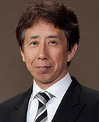 Mitsuo Kawato received a B.S. degree in physics from Tokyo University in 1976 and M.E. and Ph.D. degrees in biophysical engineering from Osaka University in 1978 and 1981, respectively. From 1981 to 1988, he was a faculty member and lecturer at Osaka University. From 1988, he was a senior researcher and then a supervisor in the ATR Auditory and Visual Perception Research Laboratories. Since 2003, he has been Director of ATR Computational Neuroscience Laboratories. Since 2004, he has been an ATR Fellow. In 2010, he became Director of ATR Brain Information Communication Research Laboratories. From 2008 to 2013 March, he served as a Research Leader of BMI/Group A, SRPBS, Japanese MEXT. In 2008, he was jointly appointed as a Research supervisor of PRESTO, JST. In 2013 November, he was jointly appointed as a Research Leader of BMI Technology/Field 3, SRPBS, and Japanese MEXT.
Abstract:
Decoded fMRI Neurofeedback (DecNef)
Mitsuo Kawato
ATR Brain Information Communication Research Laboratory Group, Kyoto, Japan
One of the most important assumptions in neuroscience is that neural activity in the brain is the cause of our mind, including consciousness. The lack of experimental tools for examining cause-and-effect relationships between brain activity and mind in systems neuroscience has severely constrained the fields progress and applicability to practical problems such as robotics or clinical issues. To fill this gap between major concepts and current technology, we developed a new technique to manipulate neural codes by applying a novel online-feedback method utilizing decoded functional magnetic resonance imaging (fMRI) signals. We call this new technique DecNef. DecNef can noninvasively induce spatial activity patterns in a specific brain area through real-time fMRI neurofeedback. More concretely, visual perceptual learning was induced without visual stimulus presentation or conscious awareness of the task (Shibata et al. 2011). DecNef can be generally described as follows. First, a decoder is constructed so that it classifies fMRI multi-voxel patterns according to some specific brain information including visual attributes, emotional states or normal and pathological states in psychiatric disorders. In the induction stage, volunteers unconsciously control their brain activity so that a desirable pattern is produced while being guided by a real-time reward signal computed as the decoder output. Thus, three important elements of fMRI DecNef are decoding of information from fMRI brain activity, real-time neurofeedback of decoded information, and unconscious reinforcement learning by participants. DecNef was recently further advanced to manipulate facial preferences or induce artificial synesthesia, or to treat psychiatric disorders. Basal ganglia was the only brain region that was activated in all of the above three studies of DecNef in the induction phase of neurofeedback training, consistent with reinforcement learning idea of DecNef.
Supported by Japanese MEXT SRPBS, BMI Technology
